Electrophoresis is one of the main molecular biology techniques, they are considered the most used technique in laboratories in everything related to nucleic acid research. Using electrophoresis, we can separate DNA and RNA fragments based on their size and electrical charge through their migration through a porous material (gel), visualize them by staining, and determine the content of nucleic acids or proteins in a sample. , thus having an estimate of its concentration.
The porous gel used in this technique acts as a molecular sieve that separates the larger molecules from the smaller ones. Smaller molecules move faster through the gel while larger ones are left behind. The mobility of the particles is also controlled by their individual electrical charge.
What is electrophoresis used for?
Electrophoresis has many practical applications, such as in forensic medicine for the identification of people who may have participated in a crime, the human genome project, the investigation of proteins and genetic mutations, since when these types of alterations exist, the Altered proteins or chromosomes, are shorter or longer appearing on an electrophoresis gel in a way different from the common and clinical diagnostic tests, allowing the timely diagnosis of diseases.
Where is electrophoresis carried out?
Electrophoresis is carried out with equipment called an electrophoresis system, which is made up of a negative charge at one end and a positive charge at the other. When inserting charged molecules, in this environment, the negative ones will go to one extreme and the positive ones to the corresponding other, eventually observing a proper migration pattern that will depend on the size and charge of the molecules.
How is gel electrophoresis carried out?
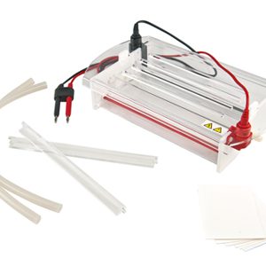
The gel used in gel electrophoresis is commonly made from agarose, which is a gelatinous substance extracted from seaweed, or also from polyacrylamide. This porous gel can be used to separate macromolecules of many different sizes. The gel is immersed in a buffer solution in an electrophoresis chamber. Tris-borate-EDTA (TBE) is commonly used as the buffer. Its main function is to control the pH of the system.
Subsequently, the samples to be analyzed are loaded into tiny wells located in the gel with the help of a pipette. Once the wells have been fully charged, a 50-150 V electrical current is applied. This causes the charged molecules present in the sample to begin migrating through the gel to the electrodes.
The speed at which each molecule travels through the gel is called its electrophoretic mobility and is primarily determined by its net charge and size. Strongly charged molecules move faster than weakly charged ones.
Once the separation is complete, the gel is stained with a dye to reveal the separation bands. Being ethidium bromide, it is a fluorescent dye commonly used in gel electrophoresis. The gel is soaked in a dilute ethidium bromide solution and then placed in a UV transilluminator to visualize the separation bands. The bands are immediately examined or photographed for future reference, as they will diffuse into the gel over time.
At Kalstein we are MANUFACTURERS and we offer you excellent electrophoresis systems at the best PRICES on the market. That is why we invite you to take a look at: HERE